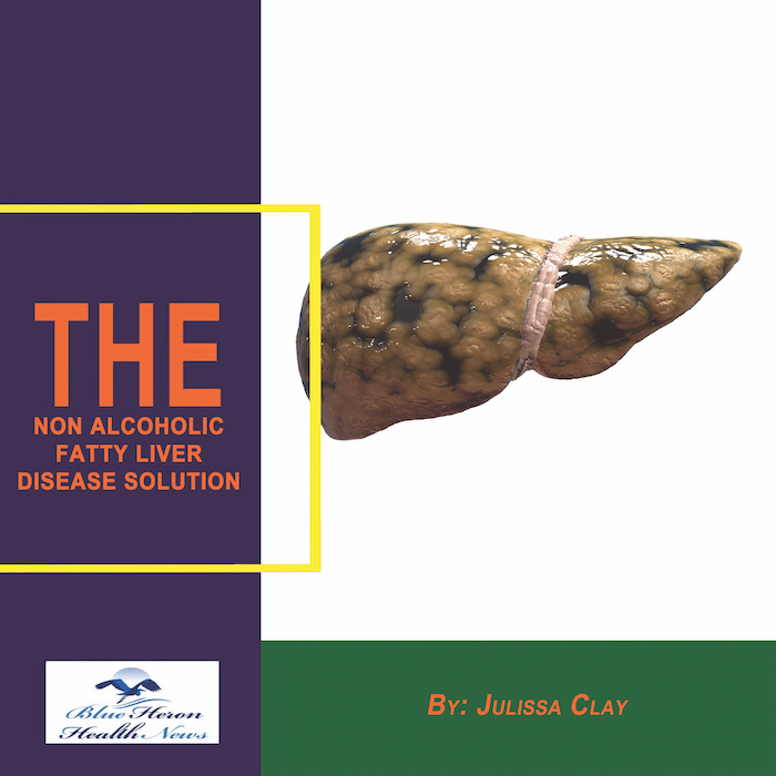
The Non Alcoholic Fatty Liver Strategy™ By Julissa Clay the program discussed in the eBook, Non Alcoholic Fatty Liver Strategy, has been designed to improve the health of your liver just by eliminating the factors and reversing the effects caused by your fatty liver. It has been made an easy-to-follow program by breaking it up into lists of recipes and stepwise instructions. Everyone can use this clinically proven program without any risk. You can claim your money back within 60 days if its results are not appealing to you.
How is a liver ultrasound used to detect fatty liver disease?
Liver Ultrasound in Detecting Fatty Liver Disease: A Detailed Explanation
A liver ultrasound is a non-invasive imaging technique commonly used to assess liver conditions, including fatty liver disease (FLD). Fatty liver disease, which can be classified into non-alcoholic fatty liver disease (NAFLD) and alcoholic fatty liver disease (AFLD), is characterized by the buildup of excess fat in liver cells. This condition can range from mild to severe and, in advanced cases, can lead to non-alcoholic steatohepatitis (NASH), fibrosis, or cirrhosis. Detecting fatty liver early is crucial for preventing disease progression, and a liver ultrasound is one of the most effective first-line diagnostic tools for this purpose.
1. The Basics of How a Liver Ultrasound Works
A liver ultrasound uses high-frequency sound waves to create images of the liver. These sound waves are sent into the body through a gel applied to the skin (usually in the upper right abdomen). The sound waves bounce off liver tissues and are received back by the ultrasound device, which creates images based on these reflections.
- Sound waves travel through tissues at different rates depending on the tissue density.
- When the sound waves hit fat deposits, they bounce back differently, creating a brighter image on the ultrasound screen (called “hyperechoic”).
- Normal liver tissue appears darker, while fat deposits make the liver appear brighter, making it easier to identify areas of fat accumulation.
2. Identifying Fatty Liver Disease Using Ultrasound
The liver ultrasound provides several critical pieces of information that can help doctors assess whether fatty liver disease is present:
A. Increased Echogenicity (Bright Liver)
- Fatty liver tissue reflects sound waves more efficiently than normal liver tissue, causing it to appear brighter (hyperechoic) on the ultrasound.
- Mild fat deposits may cause slight brightness, while moderate to severe fatty liver results in a very bright liver image.
- The more fat that accumulates in the liver, the brighter the liver will appear on the ultrasound.
B. Blurring of Liver Vasculature
- The presence of excess fat in the liver may cause blurring or loss of visibility of the blood vessels in the liver. Normally, these blood vessels can be seen clearly on the ultrasound, but fat accumulation can obscure their details.
C. Attenuation of Sound Waves
- Fatty liver tissues absorb sound waves, which causes less penetration of the sound waves into the liver. As a result, the deeper parts of the liver may not be as clearly visualized, and this phenomenon is known as attenuation. This can be a clue that fat has accumulated in the liver, especially in cases of advanced fatty liver disease.
D. Hepatomegaly (Enlarged Liver)
- In cases where fatty liver disease is present, the liver may enlarge as it becomes overloaded with fat. This enlargement is known as hepatomegaly, and it may be visible on the ultrasound image. An enlarged liver may also suggest more severe fat accumulation or liver inflammation.
3. Liver Ultrasound in Fatty Liver Disease Diagnosis
-
Detection of Fat Accumulation:
Liver ultrasound is particularly effective in detecting moderate to severe fat accumulation in the liver, which can be visible even with as little as 30% fat infiltration. However, for mild fatty liver (less than 10-20% fat), ultrasound sensitivity may be lower, meaning it might miss early-stage disease. -
Fatty Liver Grading:
Based on the ultrasound appearance, the liver can be categorized into several grades of fatty infiltration:- Grade 1 (Mild): Slight increase in liver echogenicity with normal visualization of the liver’s blood vessels.
- Grade 2 (Moderate): More pronounced brightness and partial obscuration of the blood vessels.
- Grade 3 (Severe): Very bright liver with significant loss of vasculature visibility.
4. Limitations of Liver Ultrasound for Fatty Liver Disease
While liver ultrasound is a valuable tool, it does have some limitations in the context of fatty liver disease:
A. Inability to Quantify Fat Content
- Liver ultrasound can show the presence of fat but cannot quantify the exact amount of fat present. This means it cannot provide precise information about the degree of fatty infiltration (e.g., how much fat percentage is in the liver).
- To assess mild fatty liver, imaging techniques like MRI or CT scans may be more precise.
B. Limited Detection of Early Fatty Liver Disease
- Ultrasound is more effective at identifying moderate to severe fatty liver disease, but it might not detect early, mild cases of fatty liver (less than 10-20% fat content). In early stages, the liver may not appear significantly different from normal on ultrasound.
C. No Ability to Distinguish Between Simple Fatty Liver and NASH
- Non-alcoholic steatohepatitis (NASH) involves liver inflammation and damage in addition to fat accumulation. While ultrasound can detect fat in the liver, it cannot differentiate simple fatty liver (NAFLD) from NASH, which requires liver biopsy or additional tests like MRI or FibroScan for further analysis.
5. Additional Tests Following Ultrasound
If fatty liver is detected by ultrasound, your healthcare provider may recommend further tests to assess the extent of liver damage and to rule out other causes of liver disease:
- Blood Tests (Liver Function Tests): Assess liver enzyme levels (e.g., ALT, AST) to check for inflammation or damage.
- Liver Biopsy: Considered the gold standard for assessing liver damage, inflammation, and fibrosis, especially if NASH is suspected.
- MRI or CT Scan: Provide more detailed imaging and can quantify fat content more accurately than ultrasound.
- FibroScan (Transient Elastography): Assesses liver stiffness to evaluate fibrosis or cirrhosis in people with fatty liver disease.
Conclusion
In summary, liver ultrasound is a non-invasive and effective diagnostic tool for detecting moderate to severe fatty liver disease. It helps identify fat accumulation by showing increased liver echogenicity, blurring of the liver vasculature, and signs of liver enlargement. While ultrasound cannot measure the exact amount of fat or differentiate between fatty liver and more severe liver conditions like NASH, it remains a key component in the initial screening and monitoring of fatty liver disease. Further tests may be required to assess the severity of the condition and guide treatment decisions.
Would you like more details on how ultrasound compares to other tests for fatty liver disease?
The Non Alcoholic Fatty Liver Strategy™ By Julissa Clay the program discussed in the eBook, Non Alcoholic Fatty Liver Strategy, has been designed to improve the health of your liver just by eliminating the factors and reversing the effects caused by your fatty liver. It has been made an easy-to-follow program by breaking it up into lists of recipes and stepwise instructions. Everyone can use this clinically proven program without any risk. You can claim your money back within 60 days if its results are not appealing to you