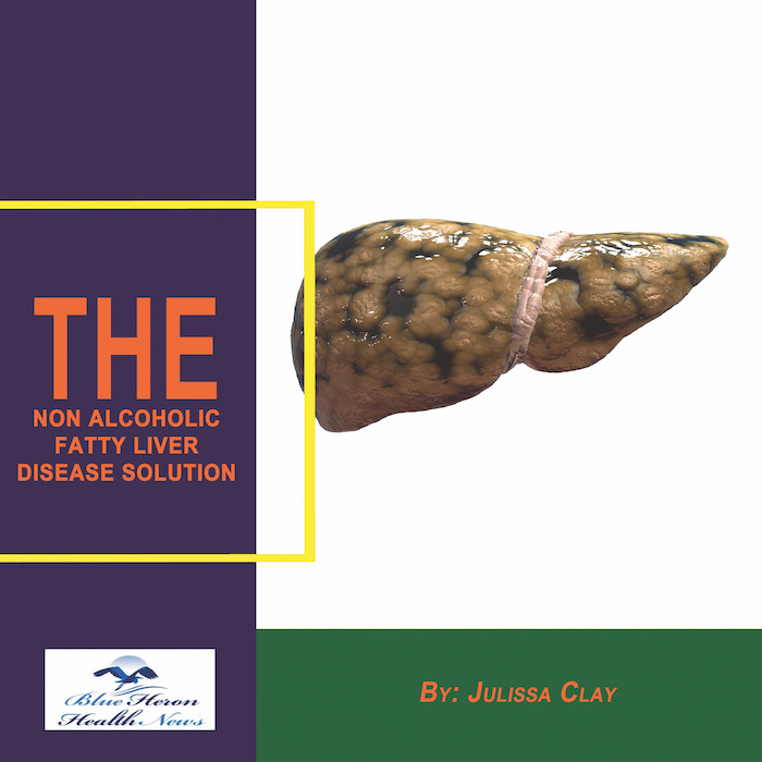
The Non Alcoholic Fatty Liver Strategy™ By Julissa Clay the program discussed in the eBook, Non Alcoholic Fatty Liver Strategy, has been designed to improve the health of your liver just by eliminating the factors and reversing the effects caused by your fatty liver. It has been made an easy-to-follow program by breaking it up into lists of recipes and stepwise instructions. Everyone can use this clinically proven program without any risk. You can claim your money back within 60 days if its results are not appealing to you.
How is fatty liver disease differentiated from other liver conditions?
Fatty liver disease is distinguished from other liver diseases by assessing the patient’s symptoms, medical history, laboratory tests, imaging tests, and in some cases, biopsy. This is how fatty liver disease (FLD) is typically distinguished from other liver diseases:
1. Medical History and Risk Factors
Fatty Liver Disease: The two most common types are non-alcoholic fatty liver disease (NAFLD) and alcoholic fatty liver disease (AFLD). NAFLD is usually associated with metabolic risk factors such as obesity, diabetes, hypertension, and dyslipidemia, while AFLD is associated with long-term alcohol consumption. A history of alcohol abuse, type 2 diabetes, or obesity will be directed toward fatty liver disease.
Other Liver Diseases: Diseases such as hepatitis B or C, cirrhosis, and liver cancer can be associated with certain risk factors such as viral infections, chronic alcoholism, or autoimmune disorders. A history of exposure to hepatitis, blood transfusions, or a family history of liver diseases can indicate other liver pathologies.
2. Symptoms
Fatty Liver Disease: Fatty liver disease is often asymptomatic in its early stages, but patients may complain of fatigue, abdominal pain, or a sensation of fullness in the upper right quadrant of the abdomen. Jaundice is uncommon in early fatty liver disease.
Other Liver Diseases: On the other hand, severe liver diseases like hepatitis or cirrhosis can have yellow skin or eyes (jaundice), swelling around the abdominal area (ascites), dark urine, and easy bruising. Severe liver diseases also bring about confusion or mental changes (hepatic encephalopathy), which are uncommon in mild fatty liver disease.
3. Laboratory Tests
Fatty Liver Disease: Mild elevations of liver enzymes (ALT, AST) may occur on blood work but typically not to the extent seen with more severe liver disease. AST/ALT ratio is helpful—typically, in fatty liver, the ratio will be less than 1, while in ALD or cirrhosis, the ratio is greater.
Other Liver Disease: Cirrhosis or hepatitis can cause greater elevations in liver enzymes, especially AST and ALT, and can also present with high bilirubin. Hepatitis B or C testing would find evidence of HBsAg, antibodies to hepatitis B or C, and viral RNA, all absent in fatty liver disease. Autoimmune conditions like autoimmune hepatitis would show an increase in autoantibodies, like ANA or ASMA.
4. Imaging Tests
Fatty Liver Disease: Ultrasound, CT scan, or MRI can identify fatty infiltrations within the liver. Ultrasound is the most commonly employed diagnostic imaging for fatty liver disease, and it may find echogenicity (brightness) of the liver. Advanced disease is assessed using a fibroScan or MRI elastography for the liver stiffness, indicating fibrosis or cirrhosis.
Other Liver Diseases: For diseases like hepatitis, cirrhosis, or cancer of the liver, imaging tests may disclose changes like hepatomegaly, nodular liver texture, ascites, or masses (e.g., liver masses). Contrast-enhanced CT or MRI will identify HCC in cirrhotic livers.
5. Liver Biopsy
Fatty Liver Disease: Liver biopsy remains the gold standard for the diagnosis and staging of fatty liver disease, particularly to differentiate between simple steatosis (fat accumulation) and non-alcoholic steatohepatitis (NASH), which involves inflammation of the liver and can progress to fibrosis and cirrhosis. The biopsy will determine the extent of fat deposition and inflammation, distinguishing it from other liver conditions.
Other Liver Diseases: The biopsy in hepatitis (viral or autoimmune), cirrhosis, or liver cancer would show different patterns of damage, including chronic inflammation, fibrosis, and necrosis in hepatitis or cirrhosis. In cirrhosis, the biopsy would show widespread scarring and nodularity of the liver.
6. Histological Features
Fatty Liver Disease: On biopsy of the liver, the hallmark of fatty liver disease is macrovesicular fat accumulation in hepatocytes. In NASH, there can be inflammation and possible fibrosis surrounding the fatty regions as well. NASH can progress to cirrhosis and liver failure but tends to be more subtle than other liver diseases.
Other Abnormalities of the Liver: Conditions like hepatitis or cirrhosis have different histological features. Hepatitis, for example, shows necrosis and inflammatory cell infiltrate in the liver tissue, while cirrhosis shows severe fibrosis, regenerative nodules, and loss of normal liver architecture. Liver cancer will show malignant tumor cells and invasion of adjacent tissues.
7. Progression and Complications
Fatty Liver Disease: Fatty liver disease, particularly NAFLD, may remain stable for a few years without progression. But it may progress to NASH, fibrosis, cirrhosis, and liver failure if not managed appropriately. NASH is more likely to develop into cirrhosis than simple fatty liver (steatosis).
Other Liver Diseases: Chronic viral hepatitis (e.g., hepatitis B or C) can lead to chronic liver disease, cirrhosis, and liver cancer. Cirrhosis from any cause (e.g., alcoholic liver disease, hemochromatosis, or autoimmune hepatitis) can lead to liver failure and susceptibility to liver cancer.
8. Ruling Out Other Conditions
Fatty Liver Disease: When diagnosing fatty liver disease, other etiologies of liver damage should be excluded, such as alcoholic liver disease, viral hepatitis, hemochromatosis, Wilson’s disease, and autoimmune hepatitis. Ruling out these diseases can be achieved through a complete evaluation of alcohol consumption, viral serology, genetic testing for hemochromatosis (e.g., HFE gene mutation), and autoimmune testing.
Other Liver Conditions: Fatty liver disease exclusion is necessary in conditions like hepatitis, cirrhosis, or liver cancer, especially where there are either metabolic risk factors or obesity. Differentiation is important, and treatment and prognosis can be very different.
Conclusion:
Fatty liver disease is distinguished from other liver conditions by a combination of medical history, clinical presentation, laboratory testing, imaging modalities, and, in some cases, liver biopsy. The most significant are the presence of fat accumulation in the liver and the absence of other markers like viral infection, autoimmune markers, or excessive alcohol consumption. Fatty liver disease in the initial stages is most often asymptomatic, while other liver conditions like hepatitis, cirrhosis, or liver cancer present with more specific manifestations and laboratory findings.
Would you like more information about the treatment or management of fatty liver disease?
Difference between fatty liver disease (FLD) and other types of liver disease blends clinical assessment, laboratory testing with blood examination, imaging examination, and even biopsy on occasions. Fatty liver disease comes in the form of non-alcoholic fatty liver disease (NAFLD) and alcoholic fatty liver disease (AFLD) and both have the potential to progress further to non-alcoholic steatohepatitis (NASH), cirrhosis, and additional liver morbidity. Therefore, fatty liver disease is distinguished from other types of liver disease as follows:
1. Clinical History
The first part of the diagnosis of fatty liver disease is a thorough clinical history, focusing on:
Alcohol consumption: To differentiate between alcoholic fatty liver disease (AFLD) and non-alcoholic fatty liver disease (NAFLD), alcohol consumption in the patient is assessed. AFLD is related to excessive alcohol consumption, while NAFLD occurs in patients with little or no alcohol consumption.
Risk factors for NAFLD: This involves obesity, type 2 diabetes, metabolic syndrome, and insulin resistance. People with these are at greater risk of getting NAFLD.
2. Physical Examination
While physical findings cannot always establish a diagnosis of fatty liver disease, there are certain findings that can suggest the presence of the disease:
Discomfort or fatigue in the upper right quadrant of the abdomen may suggest fatty liver disease.
Obesity, especially visceral fat (abdominal fat), is typically a high indicator for NAFLD.
Liver failure symptoms such as jaundice (yellowing of the skin or eyes), ascites (fluid buildup in the abdomen), or hepatomegaly (swollen liver) may be more indicative of advanced liver disease like cirrhosis rather than plain fatty liver.
3. Blood Tests
Blood tests play a significant role in distinguishing fatty liver disease from other liver conditions. Key tests are:
Liver enzymes: Raised alanine aminotransferase (ALT) and aspartate aminotransferase (AST) are characteristic of fatty liver disease, but these can also be raised in other liver conditions. In NAFLD, ALT is typically larger than AST (AST:ALT ratio <1), but in alcoholic liver disease or cirrhosis, AST is larger than ALT (AST:ALT ratio >1).
Bilirubin: Raised bilirubin is more suggestive of obstructive jaundice, hepatitis, or cirrhosis, not NAFLD.
Albumin and coagulation studies (e.g., prothrombin time): These are employed to assess the functioning of the liver. Abnormal values may point to damage to the liver, i.e., more than just simple NAFLD, i.e., cirrhosis or chronic hepatitis.
Lipid profile: Cholesterol and triglycerides are raised in NAFLD, which is typically part of metabolic syndrome.
Hepatitis markers: Rule out viral hepatitis (hepatitis B or C) by laboratory blood tests because these diseases may produce similar liver enzyme abnormalities but require different treatment.
4. Imaging Studies
Invasive imaging is commonly used to assess liver fat content and structure. These include:
Ultrasound: It is a first-line imaging agent for the diagnosis of liver fatty infiltration. While fatty liver will typically appear as a bright liver on ultrasound, the sign is not specific to fatty liver and can be seen in hepatitis or cirrhosis.
CT scan or MRI: More sensitive imaging tests, such as MRI proton density fat fraction (PDFF) or magnetic resonance elastography (MRE), are more sensitive to quantify liver fat and liver stiffness, which can indicate fibrosis (liver scarring).
Elastography: It assesses liver stiffness with ultrasound and can be applied to assess fibrosis or cirrhosis that may result from long-standing fatty liver disease.
5. Liver Biopsy
Although routine liver biopsy is not the practice, it remains the gold standard of investigation for determining the grade of liver injury, particularly where diagnosis remains in doubt. Although a biopsy distinguishes NAFLD from conditions such as NASH which also involve liver damage and inflammation, cirrhosis can be ruled out as well as other liver pathologies such as autoimmune hepatitis or hemochromatosis.
Steatosis (deposition of fat) without inflammation is characteristic of NAFLD, whereas NASH has fat with liver cell damage and inflammation.
Staging of fibrosis is required to determine whether fatty liver has progressed to cirrhosis, a condition requiring more aggressive treatment.
6. Differentiating Other Liver Diseases
Fatty liver disease may be confused with other liver diseases, but there are differentiating features:
Viral Hepatitis: Hepatitis B or C can simulate similar raised liver enzymes (AST, ALT) but is usually also accompanied by hepatitis markers (e.g., anti-HCV, HBV DNA), not present in fatty liver disease. Ultrasound or biopsy will distinguish between them.
Alcoholic Liver Disease: Care should be taken to distinguish AFLD from NAFLD. A history of excessive alcohol consumption (>30g/day in men and >20g/day in women) is crucial for the diagnosis of AFLD. NAFLD occurs in subjects who consume minimal or no alcohol.
Autoimmune Hepatitis: The disease may mimic fatty liver disease, but it is typically accompanied by increased immunoglobulins (IgG) and positive autoantibodies (e.g., ANA, SMA). Biopsy or laboratory tests will distinguish them.
Hemochromatosis: Iron accumulation in hemochromatosis can lead to liver injury analogous to that caused by fatty liver disease. Hemochromatosis is distinguished from fatty liver by iron studies (serum ferritin and transferrin saturation).
Wilson’s Disease: Copper accumulation causes fatty liver and cirrhosis. Diagnosis is established by quantitation of ceruloplasmin levels and 24-hour urine copper excretion.
7. Risk Stratification
Other investigations such as NAFLD fibrosis score, FIB-4 index, or AST-to-platelet ratio index (APRI) are used in patients who are suspected to have fatty liver disease to determine the likelihood of advanced fibrosis or cirrhosis. The scores are based on a combination of platelet count, liver enzymes, and other clinical variables to gauge the extent of liver injury.
Conclusion
The differentiation of fatty liver disease from other liver conditions involves a combination of clinical assessment, laboratory tests, imaging studies, and in certain instances, liver biopsy. Careful consideration of alcohol consumption, underlying conditions (e.g., diabetes, obesity), and utilization of diagnostic equipment like ultrasound or liver enzyme testing distinguishes NAFLD from alcoholic liver disease, viral hepatitis, and other liver conditions. Early detection and follow-up of liver function and staging of fibrosis are required to manage fatty liver disease and prevent its progression to more severe liver diseases like cirrhosis.
Would you prefer additional information about the management of fatty liver disease or clarification in terms of any specific tests?
The Non Alcoholic Fatty Liver Strategy™ By Julissa Clay the program discussed in the eBook, Non Alcoholic Fatty Liver Strategy, has been designed to improve the health of your liver just by eliminating the factors and reversing the effects caused by your fatty liver. It has been made an easy-to-follow program by breaking it up into lists of recipes and stepwise instructions. Everyone can use this clinically proven program without any risk. You can claim your money back within 60 days if its results are not appealing to you